A colored image of a fish's insides, in purple, blue, red and green

ScanAllFish/Adam Summers

STANDARDS

NGSS: Core Idea: PS3.A

CCSS: Writing: 4

TEKS: Science: 3.6A; 4.6A; 5.6A; 6.3C; ELA: 3.13A; 4.13A; 5.13A; 6.12A

Skeleton Scanner

Biologist Adam Summers reveals the insides of fish

As you read, think about what scientists can learn from looking at the insides of animals like fish.

Courtesy of Kathy Ballard

Adam Summers

Adam Summers is a marine biologist at the University of Washington. As part of his job, he investigates questions about sea creatures, like: How can some fish attach to rocks or burrow in the sand? What can armored fish teach us about protecting humans?

To answer those questions, scientists need to understand the physical structures of fish. That’s why for the past three decades, Summers has been creating 3-D images of fish skeletons. 

To make them, he uses a machine called a CT scanner (see Inside a CT Scanner, below). Summers posts the images online for researchers to study. 

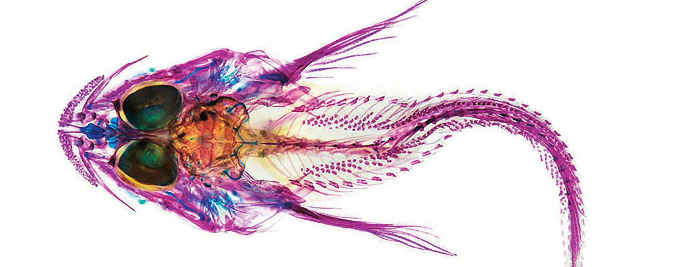
Summers has also turned fish skeleton images into vivid works of art. For these pictures, he uses a different process that involves staining the fish’s bones with colorful dyes and photographing them against a bright light. 

Summers recently spoke with SuperScience about his fish-scanning process.

Adam Summers a marine biologist. He works at the University of Washington. Summers investigates questions about sea creatures. One thing he wants to learn is how some fish attach to rocks. He wants to learn how other fish burrow in the sand. He also wants to find out what armored fish can teach us about protecting people.

How can Summers answer those questions? He needs to know more about fish bodies. That’s why he’s been making 3-D images of fish skeletons. He’s done this for the past 30 years. He makes the images using a machine called a CT scanner (see Inside a CT Scanner, below). Summers posts the images online. Other scientists can study them. 

Summers has done something else with fish skeleton images. He’s turned them into art. To make them, he stains the fish’s bones with colorful dyes. Then he photographs them. 

Summers recently spoke with SuperScience. He talked about his fish-scanning process.

What made you decide to start scanning fish?

I study biomechanics, the structure, function, and movement of living things. In 1992, a group donated a CT scanner to our lab. This machine uses invisible waves of energy called X-rays to take pictures of structures inside the body. A computer combines the images to create a 3-D picture. 

I scanned fish one at a time and enjoyed looking at the data. When I shared the pictures, people asked to scan their favorite fish. Eventually I decided to try to scan them all. So far, we have scanned over 4,000 species!

I study biomechanics. It’s the structure, function, and movement of living things. Our lab got a CT scanner in 1992. This machine uses invisible waves of energy. They’re called X-rays. They allow us to take pictures inside the body. A computer put the images together. That creates a 3-D picture.

I scanned fish one at a time. I shared the pictures. People asked me to scan their favorite fish. Eventually, I decided to scan them all. We’ve scanned more than 4,000 species so far!

How does the scanning process work?

We borrow preserved fish specimens from museums. When they come to the lab, we take them out of the plastic bags they are shipped in. We wrap several fish up together in cloth soaked with a substance called ethanol. This keeps them from drying out in the machine. Then we pop the wrapped up mass into a plastic cylinder. The cylinder goes into the CT scanner, and all the fish are scanned at the same time.

We borrow fish specimens from museums. We wrap several fish up together in cloth. It’s soaked with ethanol. This chemical keeps them from drying out in the machine. We put the wrapped fish into a container. It goes into the CT scanner. All the fish are scanned at the same time.

What new things have the scans revealed?

People have used the data for hundreds of projects. We learn all sorts of things: the anatomy of new species, how bones fit together, which fish have really dense or large bones, and how scales and bones interact.

People have used the data for hundreds of projects. We learn all sorts of things. One thing is the structure of fish bodies. We learn how fish’s bones fit together. We find out which fish have hard or large bones. And we learn how scales and bones interact.

How can the images help scientists?

We have described new species of fish, and that has been really special. It is so neat to be involved in expanding our understanding of biodiversity, Earth’s variety of living things.

We have found new species of fish. That has been really special. It’s so neat to add to our growing knowledge of biodiversity, the variety of livings on Earth.

What do you love most about your job?

I get up every morning and get to ask questions that interest me. I love finding the edges of what we know and pushing those boundaries a little further out.

I get up every morning and get to ask questions that interest me. I love finding the edges of what we know. And I love pushing those limits a little further.

Skills Sheets (3)
Skills Sheets (3)
Skills Sheets (3)
Lesson Plan (2)
Lesson Plan (2)
Leveled Articles (1)
Text-to-Speech